Where Is The Cilia Located In An Animal Cell
This folio content
1. Cilium
2. Flagellum
3. Construction
4. Movement
5. Germination
Microtubules are cytoskeleton components with important functions in jail cell physiology. The microtubule scaffold of the cytoplasm is highly plastic thank you to the polymerization and depolymerization capacity of microtubules. Notwithstanding, not all jail cell microtubules are under this shrinking or growing stages. Cilia, flagella and centrioles/basal bodies are cellular structures containing very stable (number and length) and highly organized microtubules. In this page we are dealing with cilia and flagella.
1. Cilium
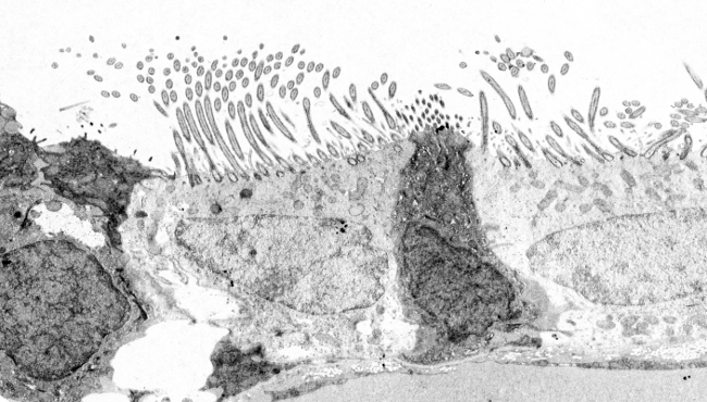
Cilia are thin and long prison cell protrusions of nearly 0.25 µm in diameter and about 10 to 15 µm in length, which can be found in animal cells and some unicellular eukaryotic species. They are ordinarily tightly packed at the gratuitous surface of epithelial cells (Figures 1 an two), such as the epithelium of the respiratory tracts, epithelium of reproductive ducts, gills of fish and bivalves, etcetera. Cilia are motile structures and their primary role is to motility the surrounding liquid, similar the mucus of the respiratory tract surface, water effectually gill epithelium, but likewise the oocyte in the female person Fallopian duct. Many unicellular organisms can move propelled by cilia, and others can utilize them for generating water swirl for catching food. Embryo nodal cilia have been implicated in initiating the left–right axis during embryonic evolution of vertebrates. Cilia movement is like beating, which impulses the liquid parallel to the jail cell surface.

Figure i. Scanning electron microscopy images showing the central culvert of a lamprey spinal string. Many cilia tin exist observed (at higher magnification in B) and small microvilli at the upmost domain of ependimal cells.

Figure 2. Transmission electron microscopy images of the respiratory epithelium. Cells with articulate cytoplasm prove many cilia in their upmost surface.
There are cilia that cannot motion, and therefore they are not intended for liquid movement. These cilia are known as master cilia. Most cells report so far (excepting crimson blood cells) bear primary cilia: oviduct cells, neurons, chondrocytes, ectoderm cells, mesenchymal cells, urinary epithelial cells, hepatocytes, and even cultured cells. Initially, master cilia were though as non-functional cilia. However, many receptor types and ion channels were found the ciliary membrane, then they were regarded as cell sensory structures. For instance, olfactory receptors are institute in cilia of their dendrites, and the external segments of rods and cones of the retin are actually modified cilia. Some receptors are more highly packed in the ciliary membrane than in other plasma membrane domains. In addition, in that location is a broad diverseness of molecules in the interior of the cilia involved in signal transduction roles. The higher surface/book ratio of a cilium makes intraciliary molecular responses more intense and efficient than if it were outside the cilium. As well chemical signal, chief cilia may discover fluid move exterior the cell and piece of work as mechanoreceptors.
2. Flagellum
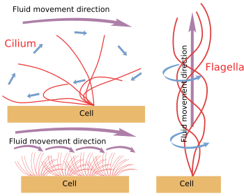
Flagella are similar to cilia, but they are much longer, nigh 150 µm long, and slightly thicker. They are quite less numerous than cilia in cells. The main function of flagella is to motion the jail cell. The flagellum motion is different from that of cilium considering the movement direction is perpendicular to the jail cell surface (not parallel), that is, the direction of the longitudinal axis of the flagellum. Flagella tin be often observed in motile cells similar unicellular organisms and sperm.
three. Construction
Cilia and flagella are complex structures containing more than 250 different proteins. Both share the aforementioned key microtubule organization and other associated proteins, altogether known as axoneme, and limited by plasma membrane (Figure 3). Also axoneme, there are many soluble molecules inside the cilia/flagella constituting the matrix. Axoneme is made up of 9 pairs of microtubules around another central pair of microtubules. This organisation can be writen equally (9 ten 2) + ii. Main cilia lack central pair of microtubules. Each microtubule of the cardinal pair is made up of xiii protofilaments, but microtubules of the peripheral pairs share some protofilaments between each other. Thus, a peripheral pair is formed by A and B microtubules. The A microtubule contains 13 protofilaments and B microtubule contains 10 or 11 protofilaments, sharing 2 or iii with A microtubule.

Figure iii. Main molecular components of cilia and flagella. In primary cilia, the central pair is absent.
The microtubule arrangement of the axoneme is the result of a scaffold of proteins. Twelve proteins have already been constitute equally constituents of the axoneme involved in maintaining microtubule organization. The neighbour peripheral microtubule pairs are connected between each other by nexin. In each pair, the A microtubule is connected past protein spokes to a central ring that contains the fundamental pair or microtubules. Dinein is a motor protein associated to the peripheral microtubules involved in the movement of cilia and flagella.

Figure 4. Ultrastructure of a cilium of an ependimal cell of the spinal string. (9+2)x2 means nine peripheral pairs and 1 cardinal pair of microtubules.
Microtubules are polymerized from basal bodies (Figures iii and 4). Basal body is fabricated upwards of nine triplet microtubules forming a cylinder (similar to centrioles). They lack a central pair of microtubules, so information technology is (9x3)+0. In each triplet, only one microtubule (A microtubule) has a consummate set of protofilaments, whereas B and C microtubules share some of them between each other. From the basal body, A and B microtubules grow and form the peripheral microtubules of the axoneme. Just above the basal body, there is region of the cilia known as transition zone containing the 9 peripheral pairs and no central pair. Immediately after the transition zone, there is the basal plate, from which the central pair of microtubules is polymerized to complete the axoneme. All microtububles accept the plus end toward the tip of the cilium/flagellum. The proximal end of the basal body (the inner one, or minus end of microtubules) is anchored to the prison cell cytoskeleton through long protein fibers chosen ciliary rootlets
Besides axoneme, cilia/flagella have other compartments. The membrane contains many receptors and channels for sensing the environment, especially in primary cilia. The fluid phase of the interior is called matrix, which helps with keeping organized the whole structure and is responsible for transducing the information gathered past membrane receptors. Other distinct areas are the basal body located at the base and the apical function of the cilium/flagellum, which contains proteins that stabilize the plus ends of microbules.
4. Movement

Effigy six. Models for cilium and flagella movement. They generate different fluid motility directions.
Due westhen cilium/flagellum are mechanically detached from the prison cell, they keep moving until the ATP shop is depleted. It means that the movement machinery is intrinsic (Figure vi). Actually, the movement is produced past sliding a peripheral pair of microtubules over the neighbour. Nexin and spoke proteins prevent the disorganization of the axoneme, but allow these movements. Dinein is the motor protein responsible for the sliding motility. The energy is provided by ATP. Dineins are anchored with its globular part to the A microtubule of 1 peripheral pair and with its tail part to the B microtubule of the adjoining pair. The molecular mechanism is like to that of the cytosolic dineins, but instead of transporting a cargo, it is moving a microtubule. For an efficient move, a coordination of the dineins of the axoneme is needed. Calcium waves in the interior of the cilia/flagella may coordinate dineins activation and alter the movement frequency when needed. It is of notice that not all dineins have to be activated at the aforementioned fourth dimension, but in synchrony.
five. Formation
Wuring differentiation, cells produce all necessary cilia and flagella for their normal physiology. It ways that all of them must exist generated from scratch. Axoneme microtubules are nucleated from A and B microtubules of the basal bodies, so 1 basal body per cilium/flagellum is needed. How multiple basal bodies are formed? There are at least 3 way of producing basal bodies: a) past using centrioles equally templates for nucleating basal bodies; b) from baggy textile known as deuterosome; c) in plants, there are singled-out protein aggregates that can nucleate basal bodies.
Numerous homo pathologies are issue of cilium/flagella flaws. They are known as ciliopathies, and include random laterality, wrong closure of the neural tube, polydactyly, cystic kidney, liver and pancreas pathologies, retina degeneration, obesity, and cognitive defects.
Bibliography
Marshall WF, Nonaka South. 2006. Cilia: tuning in to the cell's antenna. Current biology. 16:R604-R614.
Satir P, Christensen ST. 2007. Overview of structure and function of mammalian cilia. Annual review of phisiology. 69:377-400.
Source: https://mmegias.webs.uvigo.es/02-english/5-celulas/ampliaciones/7-cilio-flagelo.php
Posted by: szaboswely1945.blogspot.com

0 Response to "Where Is The Cilia Located In An Animal Cell"
Post a Comment